Saturday, 19 June 2021

The Double-Diamond Model of Design

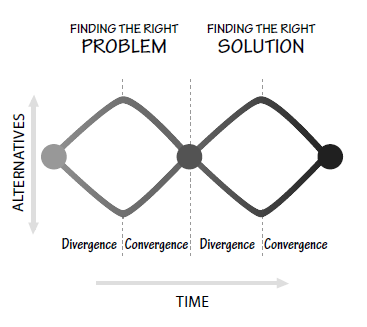
Designers often start by questioning the problem given to them: they expand the scope of the problem, diverging to examine all the fundamental issues that underlie it. Then they converge upon a single problem statement. During the solution phase of their studies, they first expand the space of possible solutions, the divergence phase. Finally, they converge upon a proposed solution (Figure 6.1). This double diverge-converge pattern was first introduced in 2005 by the British Design Council, which called it the double-diamond design process model. 
The Double-Diamond Model of Design. Start with an idea, and through the initial design research, expand the thinking to explore the fundamental issues. Only then is it time to converge upon the real, underlying problem. Similarly, use design research tools to explore a wide variety of solutions before converging upon one. (Slightly modified from the work of the British Design Council, 2005.)

The Design Council divided the design process into four stages: “discover” and “define”—for the divergence and convergence phases of finding the right problem, and “develop” and “deliver”—for the divergence and convergence phases of finding the right solution. The double diverge-converge process is quite effective at freeing designers from unnecessary restrictions to the problem and solution spaces. But you can sympathize with a product manager who, having given the designers a problem to solve, finds them questioning the assignment and insisting on travelling all over the world to seek deeper understanding. Even when the designers start focusing upon the problem, they do not seem to make progress, but instead develop a wide variety of ideas and thoughts, many only half-formed, many clearly impractical. All this can be rather unsettling to the product manager who, concerned about meeting the schedule, wants to see immediate convergence. 

To add to the frustration of the product manager, as the designers start to converge upon a solution, they may realize that they have inappropriately formulated the problem, so the entire process must be repeated (although it can go more quickly this time). This repeated divergence and convergence is important in properly determining the right problem to be solved and then the best way to solve it. It looks chaotic and ill-structured, but it actually follows well-established principles and procedures. How does the product manager keep the entire team on schedule despite the apparently random and divergent methods of designers? Encourage their free exploration, but hold them to the schedule (and budget) constraints. There is nothing like a firm deadline to get creative minds to reach convergence. 


Extracted from The Design of Everyday Things by Don Norman

Thursday, 9 January 2020

Researchers produce first laser ultrasound images of humans

Technique may help remotely image and assess health of infants, burn victims, and accident survivors in hard-to-reach places.

For most people, getting an ultrasound is a relatively easy procedure: As a technician gently presses a probe against a patient’s skin, sound waves generated by the probe travel through the skin, bouncing off muscle, fat, and other soft tissues before reflecting back to the probe, which detects and translates the waves into an image of what lies beneath.

Conventional ultrasound doesn’t expose patients to harmful radiation as X-ray and CT scanners do, and it’s generally noninvasive. But it does require contact with a patient’s body, and as such, may be limiting in situations where clinicians might want to image patients who don’t tolerate the probe well, such as babies, burn victims, or other patients with sensitive skin. Furthermore, ultrasound probe contact induces significant image variability, which is a major challenge in modern ultrasound imaging.

Now, MIT engineers have come up with an alternative to conventional ultrasound that doesn’t require contact with the body to see inside a patient. The new laser ultrasound technique leverages an eye- and skin-safe laser system to remotely image the inside of a person. When trained on a patient’s skin, one laser remotely generates sound waves that bounce through the body. A second laser remotely detects the reflected waves, which researchers then translate into an image similar to conventional ultrasound.

In a paper published today by Nature in the journal Light: Science and Applications, the team reports generating the first laser ultrasound images in humans. The researchers scanned the forearms of several volunteers and observed common tissue features such as muscle, fat, and bone, down to about 6 centimetres below the skin. These images, comparable to conventional ultrasound, were produced using remote lasers focused on a volunteer from half a meter away.

“We’re at the beginning of what we could do with laser ultrasound,” says Brian W. Anthony, a principal research scientist in MIT’s Department of Mechanical Engineering and Institute for Medical Engineering and Science (TIMES), a senior author on the paper. “Imagine we get to a point where we can do everything ultrasound can do now, but at a distance. This gives you a whole new way of seeing organs inside the body and determining properties of deep tissue, without making contact with the patient.”

Early concepts for non contact laser ultrasound for medical imaging originated from a Lincoln Laboratory program established by Rob Haupt of the Active Optical Systems Group and Chuck Wynn of the Advanced Capabilities and Technologies Group, who are co-authors on the new paper along with Matthew Johnson. From there, the research grew via collaboration with Anthony and his students, Xiang (Shawn) Zhang, who is now an MIT postdoc and is the paper’s first author, and recent doctoral graduate Jonathan Fincke, who is also a co-author. The project combined the Lincoln Laboratory researchers’ expertise in laser and optical systems with the Anthony group's experience with advanced ultrasound systems and medical image reconstruction.

Yelling into a canyon — with a flashlight

In recent years, researchers have explored laser-based methods in ultrasound excitation in a field known as photo acoustics. Instead of directly sending sound waves into the body, the idea is to send in light, in the form of a pulsed laser tuned at a particular wavelength, that penetrates the skin and is absorbed by blood vessels.

The blood vessels rapidly expand and relax — instantly heated by a laser pulse then rapidly cooled by the body back to their original size — only to be struck again by another light pulse. The resulting mechanical vibrations generate sound waves that travel back up, where they can be detected by transducers placed on the skin and translated into a photo acoustic image.

While photo acoustics uses lasers to remotely probe internal structures, the technique still requires a detector in direct contact with the body in order to pick up the sound waves. What’s more, light can only travel a short distance into the skin before fading away. As a result, other researchers have used photo acoustics to image blood vessels just beneath the skin, but not much deeper.

Since sound waves travel further into the body than light, Zhang, Anthony, and their colleagues looked for a way to convert a laser beam’s light into sound waves at the surface of the skin, in order to image deeper in the body. 

Based on their research, the team selected 1,550-nano meter lasers, a wavelength which is highly absorbed by water (and is eye- and skin-safe with a large safety margin).  As skin is essentially composed of water, the team reasoned that it should efficiently absorb this light, and heat up and expand in response. As it oscillates back to its normal state, the skin itself should produce sound waves that propagate through the body.

The researchers tested this idea with a laser setup, using one pulsed laser set at 1,550 nano-meters to generate sound waves, and a second continuous laser, tuned to the same wavelength, to remotely detect reflected sound waves.  This second laser is a sensitive motion detector that measures vibrations on the skin surface caused by the sound waves bouncing off muscle, fat, and other tissues. Skin surface motion, generated by the reflected sound waves, causes a change in the laser’s frequency, which can be measured. By mechanically scanning the lasers over the body, scientists can acquire data at different locations and generate an image of the region.

“It’s like we’re constantly yelling into the Grand Canyon while walking along the wall and listening at different locations,” Anthony says. “That then gives you enough data to figure out the geometry of all the things inside that the waves bounced against — and the yelling is done with a flashlight.”

In-home imaging

The researchers first used the new setup to image metal objects embedded in a gelatin mold roughly resembling skin’s water content. They imaged the same gelatin using a commercial ultrasound probe and found both images were encouragingly similar. They moved on to image excised animal tissue — in this case, pig skin — where they found laser ultrasound could distinguish subtler features, such as the boundary between muscle, fat, and bone.

Finally, the team carried out the first laser ultrasound experiments in humans, using a protocol that was approved by the MIT Committee on the Use of Humans as Experimental Subjects. After scanning the forearms of several healthy volunteers, the researchers produced the first fully non contact laser ultrasound images of a human. The fat, muscle, and tissue boundaries are clearly visible and comparable to images generated using commercial, contact-based ultrasound probes.

The researchers plan to improve their technique, and they are looking for ways to boost the system’s performance to resolve fine features in the tissue. They are also looking to hone the detection laser’s capabilities. Further down the road, they hope to miniaturise the laser setup, so that laser ultrasound might one day be deployed as a portable device.

“I can imagine a scenario where you’re able to do this in the home,” Anthony says. “When I get up in the morning, I can get an image of my thyroid or arteries, and can have in-home physiological imaging inside of my body. You could imagine deploying this in the ambient environment to get an understanding of your internal state.” 

This research was supported in part by the MIT Lincoln Laboratory Biomedical Line Program for the United States Air Force and by the U.S. Army Medical Research and Material Command's Military Operational Medicine Research Program.
Content credits:Jennifer Chu  http://news.mit.edu/

Saturday, 3 August 2019

We are now back in action


Sunday, 5 August 2018

BUILTROBOTICS

BUILTROBOTICS -Building the future of construction.


Are you an employer who thinks about labor shortage. Get to know about a company who made use of sensors to made smart machines to meet the requirements of construction and earthmoving. They made use of sensors and actuators to make the heavy ones smart.


To Know More visit: Built Robotics

Sunday, 1 April 2018

What’s on Your Mind? Bosses Are Using Artificial Intelligence to Find Out

AI tools give companies instant insights from employee surveys that once took months to process

By 

Wednesday, 28 March 2018

Cosmonauts Prepare to Bring the 'Internet of Animals' Online


The Icarus project will track tens of thousands of animals from the International Space Station

                                                                    By Prachi Patel


An ambitious project to keep an eye on thousands of animals and birds from space in a sort of “Internet of Animals” is getting ready to kick off.
In February, German researchers sent three large 200-kilogram antennas to the International Space Station (ISS) on a Soyuz rocket. The antennas joined a computer that had been sent up in October. These pieces will be the ears and brain of ICARUS, short for International Cooperation for Animal Research Using Space, an initiative funded by the Russian and German space agencies to track the movement of the smallest animals—birds, turtles, fish, and even insects—and tap into swarm intelligence.
Icarus researchers will outfit animals with tiny sensor-laden tags that will send their data to the computer aboard the ISS, which will clean it up and beam it down to a smart, central database. “Technically, it’s an Internet of Things via satellite,” says project leader Martin Wikelski at the Max Planck Institute for Ornithology.


Remote monitoring of animals isn’t new. It has revealed songbird migration patterns, allowed scientists to map ocean floors, and helped wildlife authorities to catch poachers. But Icarus is animal-tracking on steroids. It will allow scientists to scan all corners of the Earth to collect astounding details on tagged animals and their environment. 
The idea is that this large amount of data will reveal global patterns in animal populations, allow scientists to precisely track movements and living conditions, and show why and how animals die—information which could help protect species. 
Animals could then, in turn, aid us. Researchers could use such data to monitor fisheries and other human food sources, unravel the origin and spread of disease like Ebola and avian flu, understand climate change, and predict natural disasters, Wikelski says. “There’s good scientific data showing that animals can anticipate earthquakes, volcanic eruptions, and tsunamis.”
The German team has made tiny tags loaded with a GPS receiver, 3D accelerometer, and temperature, humidity, pressure, altitude, and heart rate sensors. The tags are also equipped with solar panels and rechargeable batteries. The smallest tags weigh 2.5 grams but the team is miniaturizing them further for songbirds and honeybees, Wikelski says.
Today’s geolocation tags burn a lot of power transmitting data via cellular networks or satellite systems. But Icarus tags use a special code-division multiple access (CDMA) coding scheme to communicate with satellites using very little power. Plus, the tags are interactive: scientists can ping them for additional data if needed. (The tags designed for fish will fall off and float to the surface in order to transmit.)
The Icarus tag is activated when it gets a signal from the orbiting computer. Then, it has two seconds to send its data up to the receiving antennas. “Large antennas are important because you can’t beat physics,” Wikelski says. “If you have a small tag on ground, you need a big antenna in space.” The on-board computer can pick up data from 120 tags at a time. It separates, parses, and cleans up the data and sends back the relevant information to Movebank, an open-source, online database the team has developed.


Wikelski and his colleagues have tested the system on the ground by outfitting larger animals with the tags and collecting data via land-based antenna. They were able to predict eruptions of Italy's Mount Etna six hours in advance by observing moving patterns of goats on the volcano’s slopes. Tagging flying foxes in Africa showed how these foxes spread seeds across vast areas. In the future, Wikelski says, the team could track the spread of diseases like avian flu or Ebola, by testing bats for antibodies that show up in their blood after disease exposure, and then figuring out where those animals have been.
The system’s real test will come in August, when the project's operational phase is scheduled to begin. For now, the Russian team is testing the computer system on the ISS. And Russian cosmonauts are training for a five-hour spacewalk in June to install and connect the antennas on the outside of the space station.
Engineering Insights

Get an Insight into the world of engineering. Get to know about the trending topics in the field of engineering.

Pages

Follow Us